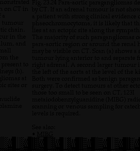
Labels:text | screenshot | black and white | black | font | document OCR: honstrated Fig. 23.24 Para-aortic paragangliomas de on CT in by CT. If an adrenal tumour is not show a a patient with strong clinical evidence tumour phaeochromocytoma, it is likely that th tic chain. lies at an ectopic site along the sympath cur in the The majority of such paragangliomas o lum, and para-aortic region or around the renal hall may be visible on CT. Scan (a) shows a om the tumour lying anterior to and separate f present to right adrenal. A second larger tumour neys (b). the left of the aorta at the level of the k liomas at Both were confirmed as benign paragan ic sites or surgery. To detect tumours at other ecto those too small to be seen on CT, 1231 nuclide metaiodobenzylguanidine (MIBG) radio lamine scanning or venous sampling for catech levels is required. See also: · MIB ...